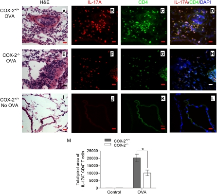
Figure 2.
Reduced Th17 cells in lung of cyclooxygenase (COX)-2−/− mice after ovalbumin (OVA) exposure. COX-2+/+ and COX-2−/− mice were sensitized with OVA in adjuvant (or given adjuvant alone). Fourteen to 21 days later, mice were exposed to inhaled OVA (or inhaled phosphate-buffered saline) for 4 consecutive days. Forty-eight hours after final OVA (or phosphate-buffered saline) exposure, lung tissue sections were stained with hematoxylin and eosin (H&E) and visualized by light microscopy (A, E, and I) (original magnification ×100). Visualization of Th17 cells in COX-2+/+ and COX-2−/− lung tissue sections was accomplished by immunofluorescence staining with phycoerythrin (PE)-labeled anti–IL-17A (B, F, and J) and fluorescein isothiocyanate (FITC)–labeled anti-CD4 (C, G, and K) antibodies. D, H, and L are merged images of anti–IL-17A and PE, anti–CD4 and FITC, and DAPI. All immunofluorescent images are shown at original magnification ×60; numerical aperture, 1.4; scale bar, 25 μm. Results are representative of three independent experiments. Multiple (n = 22–29) randomly selected regions (1,300 × 1,030 pixel2 = 350 × 277 μm2) from each lung section were counted and quantified by a masked observer using MetaMorph software (M); *P < 0.05 versus COX-2+/+.

