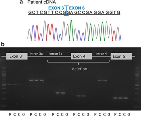
Figure 4.
(a) Electropherograms of LRP5 cDNA amplicons from patient 4 revealed the loss of exons 4 and 5. (b) PCR-amplified fragments from genomic DNA of patient 4 and two controls visualized on a 2% ethidium bromide gel. All studied amplicons; exons 3, 4 and 5, areas of intron 3 (a: 2735–3671 nucleotides and b: 4198–4503 nucleotides downstream of exon 3) and intron 4 (607–1213 nucleotides downstream of exon 4) can be detected in both control samples (C) and are clean in water samples (0) serving as negative controls. PCR of the patient's (P) genomic DNA was repeatedly unsuccessful in the areas of intron 3b, exon 4 and intron 4, suggesting a homozygous deletion involving exon 4 and adjacent intronic parts. Exon 5 and at least 10 nucleotides of the adjoining introns could be sequenced from both patient and control samples.

