
Figure 2.
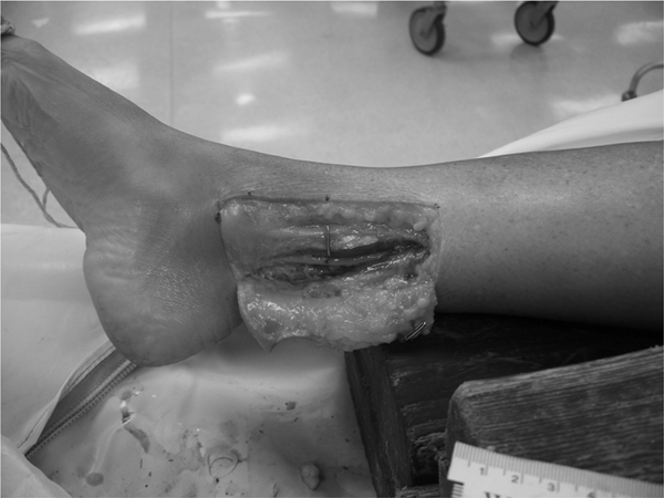
Before dye injection, location of window for dissection was identified and marked starting just proximal to the distal tip of the medial malleolus. The skin flap was incised and the neurovascular bundle was separated in order to free the tibial nerve from all adjacent tissue. This was necessary in order to visualize and measure the nerve. The injection site was marked with a coloured marker as well as a T-pin.