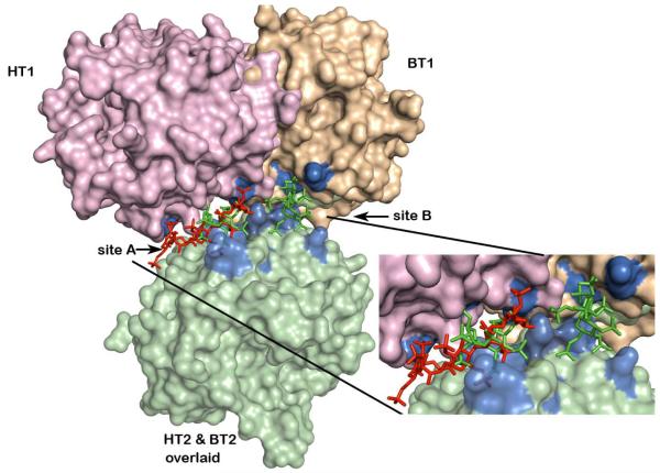
Figure 4.
Differences in monomer contacts between HT-LMWH complex and BT-SOS complex dimers. Monomer AB (HT2) of the AB-GH dimer of the HT-heparin complex was superposed on monomer XD (BT2) of the AB-CD dimer of the BT-SOS complex (light green surface). The relative positions after this superposition of the monomer mates (GH (HT1) in the HT-heparin complex and AB (BT1) in the BT-SOS complex) in each of the dimers are colored pink and wheat respectively. LMWH (red stick) bound to the HT-LMWH dimer and SOS (green stick) bound to sites 1 and 2 of the BT-SOS complex, after superposition of HT2 and BT2 monomers. Both ligands occupy the extended exosite II (residues surface highlighted in blue) shown in exploded view.

