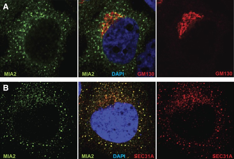
Fig. 3.
MIA2 localizes to ER exit sites in HuH-7 cells. A: Confocal imaging of HuH-7 cells costained with anti-MIA2 (green) and anti-GM130 (red) revealed that the perinuclear anti-MIA2 staining was adjacent to but not overlapping with GM130 signal. B: In this confocal image, costaining with anti-MIA2 (green) and anti-Sec31A (red) showed extensive colocalization of MIA2 with the COPII protein, Sec31. Nuclei were stained with 4’,6-diamidino-2-phenylindole (DAPI, blue).

