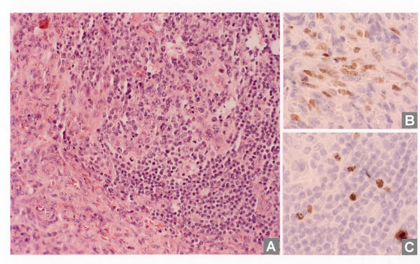
Figure 1.
Microscopic morphology of representative lymph node sections and immunohistochemical detection of HHV8 infection. A: Histology of the axillary lymph node showing proliferation of spindle cells (lower left) and partly involuted germinal center, surrounded by small lymphocytes (upper right). B: HHV8 positive spindle cells. C: HHV8 positive lymphocytes in the follicular mantle zone.

