FIGURE 1.
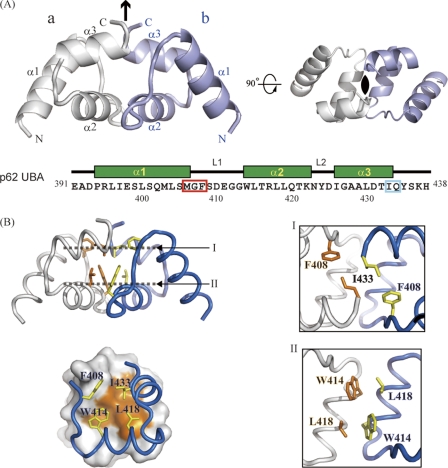
Crystal structure of p62 UBA dimer. A, ribbon representation of the crystal structure of the p62 UBA dimer. Noncrystallographic C2 symmetry axis is shown as a black arrow and a black oval. Each monomer unit is colored in white or light blue. The amino acid sequence with secondary structure indicated is shown below the structures. In the sequence, the MGF motif and the residues corresponding to di-leucine motif in canonical UBA domains are indicated by the red and cyan box, respectively. B, residues at the dimerization interface of the p62 UBA dimer are shown as stick models. Interactions in the dimer interface are shown in the right panel. The locations of planes I and II are indicated in the left panel, top. Each plane is the top view of the structure.

