FIGURE 6.
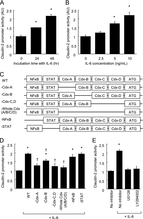
IL-6 enhances claudin-2 promoter activity in a Cdx binding site-dependent manner. A and B, claudin-2 reporter gene plasmids were transfected to Caco-2 cells. Luciferase activity was measured in the cell monolayers incubated in the absence or presence of IL-6 (10 ng/ml) for 24 and 48 h (A). Luciferase activity was measured in Caco-2 cell monolayers incubated with varying concentrations of IL-6 (0–10 ng/ml) for 48 h (B). *, p < 0.05 relative to control treatment. C, schematic drawings of transcriptional binding sites in the WT-claudin-2 promoter and deletion mutants. D, WT-claudin-2 reporter gene plasmids and the deletion mutant plasmids were transfected to Caco-2 cells. Luciferase activity was measured in the cell monolayers incubated in the absence or presence of IL-6 (10 ng/ml) for 48 h. *, p < 0.05 relative to control treatment. †, p < 0.05 relative to WT-claudin-2 with IL-6. E, WT-claudin-2 reporter gene plasmid was transfected to Caco-2 cells. Luciferase activity was measured in the cell monolayers incubated in IL-6-free medium or in medium containing 10 ng/ml IL-6 in the absence or presence of signaling inhibitors (U0126, a MEK inhibitor; LY294002, a PI3K inhibitor) for 48 h. *, p < 0.05 relative to control treatment. Values represent the mean ± S.E. (n = 4).

