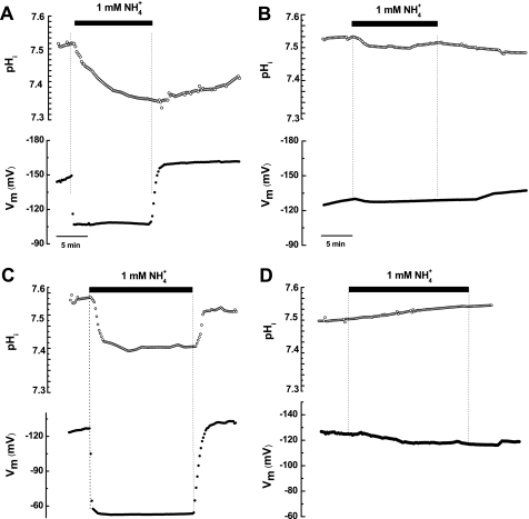
FIGURE 3.
Ammonium induces cytoplasm acidification in oocytes expressing PvAMT1;1. A, acidification of the cytoplasm (top) and membrane potential depolarization (bottom) caused by exposure of an oocyte expressing PvMT1;1 to 1 mm NH4+ at pH 7.0. Upon ammonium removal, both cytoplasm pH and oocyte membrane potential recovered to their initial levels, with the former taking more than 10 min. B, a control DEPC/H2O-injected oocyte exposed to the same conditions showed smaller acidification (top) and smaller change in membrane potential (bottom). C, acidification (top) and membrane depolarization (bottom) caused by 1 mm NH4+ in an oocyte expressing PvMT1;1 exposed to pH 5.5. Observe the faster changes in both parameters when compare with those recorded at pH 7.0 (see A). D, changes in cytoplasmic pH (top) and membrane potential (bottom) caused by 1 mm NH4+ in a control oocyte exposed to pH 5.5. Observe the slight alkalinization upon exposure to ammonium.

