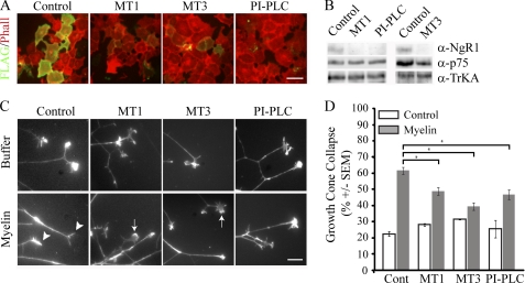
FIGURE 4.
MT1-MMP- and MT3-MMP-dependent NgR1 shedding attenuates myelin-dependent growth cone collapse. A, cell-surface FLAG and rhodamine-phalloidin (Phall) staining of COS-7 cells transfected with FLAG-NgR1 and treated with 0.75 μm MT1-MMP (MT1), 1.5 μm MT3-MMP (MT3), or 1 unit/ml PI-PLC for 3 h. Scale bar = 10 μm. B, Western blotting of biotinylated cell-surface proteins from P5 DRG neurons treated with recombinant MT-MMPs or PI-PLC. C and D, growth cones of P5 rat DRG explants treated with buffer or myelin extract. Explants were pretreated for 3 h with control buffer or MT1-MMP (0.75 μm), MT3-MMP (1.5 μm), or PI-PLC (1 unit/ml). Growth cones were stained with rhodamine-phalloidin (C; scale bar = 10 μm) and quantified for growth cone collapse (D). Arrows and arrowheads indicate examples of spread and collapsed growth cones, respectively. Growth cone collapse quantification is from three separate experiments performed in duplicate. All samples were blinded prior to quantification. *, p < 0.05 by one-way ANOVA with Tukey's post hoc test.

