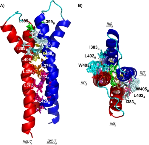
FIGURE 7.
TM6/TM7 dimerization interface in 10 structures of MT7-hM1 dimer complex. hM1A and hM1B are displayed in red and blue schematics, respectively. The transverse view (A) and extracellular view (B) are on the left and right, respectively. Three contacts at the interface are supported by the same residues on both protomers: residues Leu-376, Leu-399, and Leu-406 are colored in orange, green, and yellow, respectively. The cluster of Ile-383 of hM1A and Leu-402 and Trp-405 of hM1B and the cluster of Ile-383 of hM1B and Leu-402 and Trp-405 of hM1A are colored in pale cyan and cyan, respectively. The cluster of Leu-372 of hM1A and Ile-413, Cys-417, and Leu-420 of hM1B is colored in hot pink, and the cluster of Leu-372 of hM1B and Ile-413, Cys-417, and Leu-420 of hM1A is colored in violet.

