FIGURE 6.
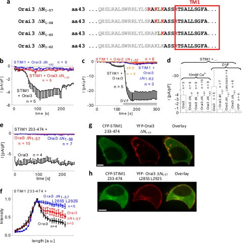
N-terminal deletions until position 57 or further significantly diminish store-operated Orai3 activation. a, amino acid sequence of Orai3 N-terminal deletion mutants. Gray letters represent amino acids within the N terminus of Orai3 that were deleted. b, time course of whole cell inward currents at −74 mV activated by passive store depletion of HEK 293 cells coexpressing CFP-STIM1 with YFP-Orai3 ΔN1–57 and YFP-Orai3 ΔN1–62 in comparison with wild-type YFP-Orai3. c, time course of whole cell inward currents at −74 mV upon perfusion with divalent cation-free (DVF) solution of HEK 293 cells expressing YFP or coexpressing CFP-STIM1 with YFP-Orai3, YFP-Orai3 ΔN1–57, or YFP-Orai3 ΔN1–62. d, block diagram comparing maximal currents in 10 mm Ca2+ of HEK cells coexpressing CFP-STIM1 and YFP-Orai3, YFP-Orai3 ΔN1–57, YFP-Orai3 ΔN1–58, YFP-Orai3 ΔN1–60, or YFP-Orai3ΔN1–62 (p < 0.05) as well as in divalent cation-free (DVF) solution of HEK 293 cells expressing YFP or coexpressing CFP-STIM1 and YFP-Orai3, YFP-Orai3 ΔN1–57, YFP-Orai3 ΔN1–57 L285S/L292S, or YFP-Orai3 ΔN1–62 (p < 0.05). e, time course of whole cell inward currents at −74 mV activated by passive store depletion of HEK 293 cells coexpressing CFP-STIM1 233–474 with YFP-Orai3 ΔN1–57 and YFP-Orai3 ΔN1–58 in comparison with wild-type YFP-Orai3 (p < 0.05). f, intensity plots representing the localization of CFP-STIM1 233–474 across the cell when coexpressed with YFP-Orai3 ΔN1–57 L285S/L292S compared with YFP-Orai3 ΔN1–57 and wild-type Orai3. g and h, localization and overlay of CFP-STIM1 233–474 and YFP-Orai3, YFP-Orai3 ΔN1–57, or YFP-Orai3 ΔN1–57 L285S/L292S. Bars in fluorescence images correspond to 5 μm. pF, picofarad. Error bars represent S.E.

