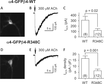
FIGURE 6.
ACh-evoked currents in neurons transfected with α4-eGFPβ4wt and α4-eGFPβ4-R348C nAChRs. The transfected neurons were identified with green fluorescence (A and D). ACh (300 μm) was puffed (0.1 s) onto voltage-clamped neurons (VH = −65 mV) and evoked inward currents (B and E). C, summary of 300 μm ACh-evoked currents (IACh) from neurons with different transfections (WT, α4-eGFPβ4wt; R348C, α4-eGFPβ4-R348C). F, summary of the density of 300 μm ACh-induced currents (IACh divided by membrane capacitance). Numbers of recorded neurons are indicated in parentheses.

