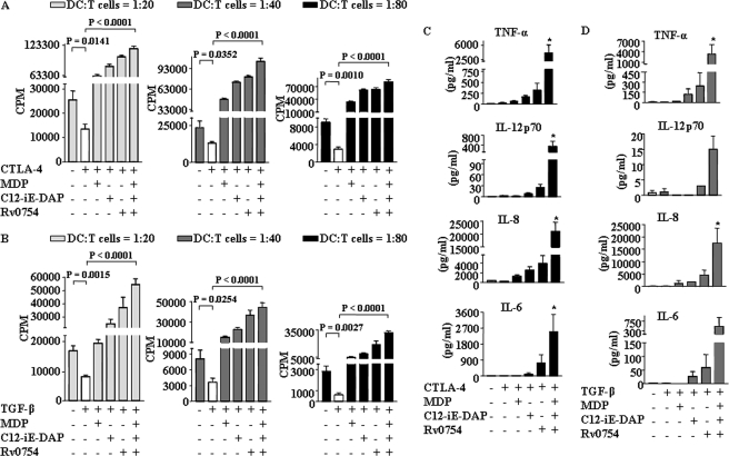
FIGURE 5.
Synergistic activation of TLR2, NOD1, and NOD2 renders human DCs to trigger strong T cell response as well as to secrete pro-inflammatory cytokines under CTLA-4- and TGF-β-induced immunosuppressive conditions. A and B, DCs were treated as indicated in Fig. 3, A–F, and were co-cultured with allogenic CD4+ T cells at different DC to T cell ratios. After 4 days of co-culture, cells were pulsed overnight with 0.5 μCi of [3H]thymidine to quantify T cell proliferation. Radioactive incorporation was expressed as counts/min (mean ± S.E. of quadruplet values). Data are presented as mean ± S.E. from four independent donors. C and D, DCs were cultured with GM-CSF and IL-4 followed by treatment with CTLA-4 (1 μg/ml) (C) or TGF-β (10 ng/ml) (D) for 6 h. DCs were further treated for 42 h with Rv0754 or C12-iE-DAP or MDP alone as well as with a combination of Rv0754, C12-iE-DAP, and MDP and secretion of IL-6, IL-8, IL-12p70, and TNF-α in cell-free culture supernatants was analyzed. *, p < 0.05 versus CTLA-4 or TGF-β.

