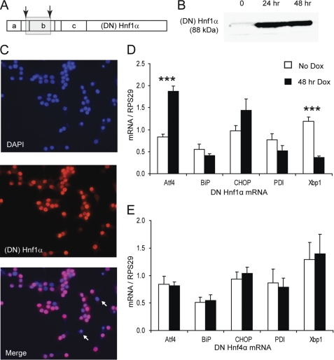
FIGURE 1.
DN HNF1α provokes an atypical ER stress response. A, map of HNF1α domain structure with positions of DN HNF1α frameshift mutations indicated by arrows. a, dimerization domain; b, DNA binding (POU-like) domain; c, homeodomain-like region. The gray box indicates the region mistranslated in the DN mutant due to the frameshifts. B, Western blot for HNF1α (4 μg of nuclear extracts/lane) after 0, 24, or 48 h of 500 ng/ml Dox. C, immunofluorescence for (DN) HNF1α after 48 h of Dox. Two non-expressing cells are marked with arrows. D, Q-PCR of ER stress genes with or without 48 h of Dox in INS DN Hnf1α cells (n = 4). E, Q-PCR of ER stress genes with or without 48 h of Dox in INS DN Hnf4α cells (n = 4). Error bars, S.E.

