FIGURE 4.
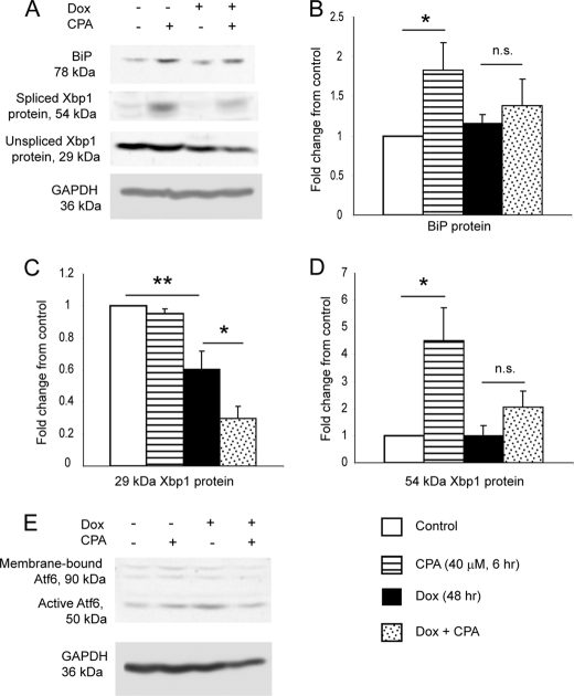
Induction of DN HNF1α prevents CPA-induced increase in BiP and 54 kDa XBP1 protein and reduces 29 kDa XBP1 protein. A, representative images of Western blots for BiP, both isoforms of XBP1, and GAPDH, after treatment with or without 48 h of Dox and 6 h of 40 μm CPA or vehicle (DMSO). B, densitometry quantification of BiP from Western blotting (n = 4). C, densitometry quantification of 29 kDa XBP1 from Western blotting (n = 4). D, densitometry quantification of 54 kDa XBP1 from Western blotting (n = 4). E, Western blot for ATF6 and GAPDH (representative image of two independent experiments). It is likely that the membrane-bound form of ATF6 is underrepresented in the image due to less efficient transfer of larger proteins to the membrane and/or less efficient extraction of membrane-bound proteins during cell lysate preparation. Numerical data are displayed as mean ± S.E. (error bars) of the -fold changes in intensity normalized to the untreated control sample. The increases (for BiP and 54 kDa XBP1) or decreases (for 29 kDa XBP1) in protein levels with CPA relative to vehicle were measured by unpaired one-tailed t test.

