FIGURE 3.
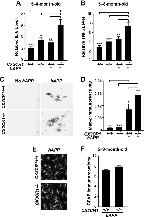
Deletion of CX3CR1 exacerbates microglial activation. A and B, quantification of levels of IL-6 (A) or TNF-α (B) mRNA in the cortex of 5–8-month-old CX3CR1+/+, hAPP/CX3CR1+/+, CX3CR1−/−, or hAPP/CX3CR1−/− mice. n = 13–19 mice/genotype. C, representative picture of Mac-2 immunostaining in the hippocampus of 5–8-month-old wild type (CX3CR1+/+), hAPP/CX3CR1+/+, CX3CR1−/−, or hAPP/CX3CR1−/− mice. D, quantification of Mac-2 immunoreactive protein in the hippocampus. n = 8–9/genotype. E, representative images of GFAP immunostaining in the hippocampus of 5–8-month-old hAPP/CX3CR1+/+ or hAPP/CX3CR1−/− mice. F, quantification of immunoreactive GFAP in the hippocampus. n = 8–10/genotype. *, p < 0.05; **, p < 0.01; ***, p < 0.001, one-way analysis of variance Tukey-Kramer post hoc analysis (A, B, and D). Values are means ± S.E.

