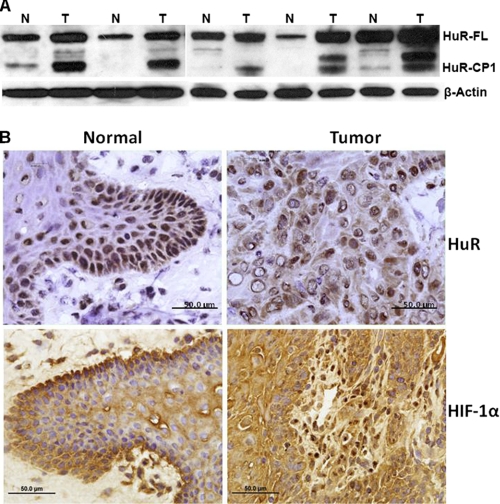
FIGURE 1.
Expression and cleavage of HuR in oral cancer tissues. A, total protein (50 μg) from the tongue tumor (T) and adjacent normal (N) tissues used for Western blot analysis is shown. The blots were probed for HuR and β-actin (used as loading control). HuR-FL indicates the 36-kDa full-length (FL) HuR; HuR-CP1 is a 24-kDa protein. B, tissue sections from representative tumors were subjected to immunohistochemistry using a primary monoclonal antibody to HuR and HIF-1α followed by peroxidase-conjugated goat anti-mouse secondary antibody. Expression of all proteins was relatively high in tumors (brown), where HuR and HIF-1α were significantly expressed in the cytoplasm. The scale bar denotes 50 μm in diameter.

