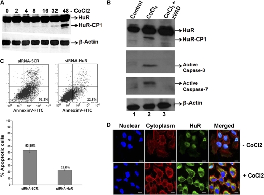
FIGURE 2.
Expression and cleavage of HuR during CoCl2-induced hypoxia. A, total protein (50 μg) from UM74B oral cancer cells was grown under 250 μm CoCl2 used for Western blot analysis. Cells were treated and harvested at the indicated time points to identify HuR cleavage. The blots were probed for HuR and β-actin (used as loading control). B, active caspases are required for the appearance of the 24-kDa HuR fragment. Cells were incubated with DMSO (first lane 1), CoCl2 (second lane), and CoCl2 + benzyloxycarbonyl-Val-Ala-Asp (zVAD; third lane) followed by Western blotting for HuR, performed as described above and probed with antibodies to active caspases-3 and -7. β-Actin serves as a loading control. C, UM74B cells treated with siRNA are described under “Experimental Procedures” in the presence of 250 μm CoCl2 for 36 h were analyzed by staining with annexin V-FITC and propidium iodide by flow cytometry. The percentage of apoptotic cells (top boxes) upon CoCl2 treatment was determined for HuR (siRNA-HuR) or control siRNA (siRNA-SCR)-treated HeLa cells. The values were normalized to control untreated cells. The graph represents the number of apoptotic cells after treatment as described in C. Values are the means ± S.E. (error bars) from three independent experiments. D, immunofluorescence detection of HuR in UM74B cells, either untreated or treated with CoCl2 as indicated. Distribution of cytoplasmic HuR (merged panel) was observed after 24 h. Blue indicates DAPI staining used to detect nuclei, red indicates β-actin staining to detect cytoplasm, and green indicates HuR immunofluorescence. The scale bar denotes 5 μm.

