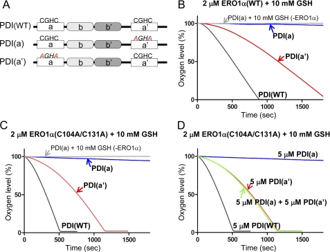
FIGURE 5.
Selective oxidation of ERO1α toward the a′ domain and the intramolecular electron relay within PDI. A, schematic representation of human PDI proteins with the -CGHC- active sites and the mutated -AGHA sites indicated. Kinetics of oxygen consumption by 2 μm wild-type ERO1α (B), or constitutively active ERO1α (C), during the reaction with 5 μm human PDI variants, as depicted in the figure, in the presence of GSH (10 mm). D, oxygen consumption by the 2 μm constitutively active ERO1α during the oxidation of 5 μm human PDI variants, including PDI(WT), PDI(a), and PDI(a′), or the mixture of PDI(a) and PDI(a′).

