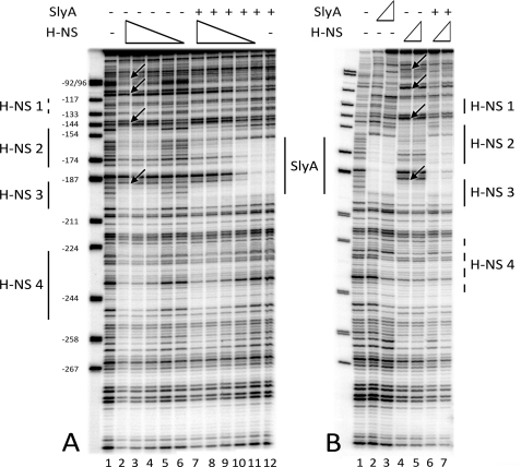
FIGURE 5.
Competition between H-NS and SlyA binding upstream of the fimB promoter. A, binding of H-NS to the fim03 fragment labeled at the fim03f end. Decreasing concentrations of H-NS were mixed with the fim03 DNA with and without SlyA in the “HNS” buffer. Lane 1, no proteins; lanes 2 and 7, 400 nm H-NS; lanes 3 and 8, 200 nm H-NS; lanes 4 and 9, 100 nm H-NS; lanes 5 and 10, 50 nm H-NS; lanes 6 and 12, 25 nm H-NS; lanes 7–12 also contained 2 μm SlyA. B, H-NS binding with and without SlyA in the “SlyA” buffer. Lane 1, no proteins; lane 2, 50 nm SlyA; lane 3, 1 μm SlyA; lane 4, 50 nm H-NS; lane 5, 100 nm H-NS; lane 6, 1 μm SlyA, and 50 nm H-NS; lane 7, 1 μm SlyA and 100 nm H-NS. Proteins were incubated for 15 min at 25 °C before treatment with DNase I. Products were analyzed on 6% denaturing polyacrylamide gels. Regions protected by H-NS and SlyA are indicated. The arrows indicate hypersensitive DNaseI cleavages on the DNA in the presence of H-NS, which are observed in the “SlyA” buffer (B) but not in the “H-NS” buffer (H-NS). The marker is pBR322 digested with MspI, and the sizes of the fragments were used to calculate their positions relative to the transcriptional start site of fimB (+1).

