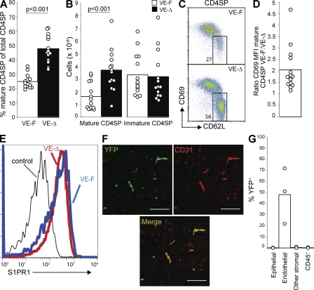
Figure 5.
LPP3 expression by endothelial cells is required to maintain low thymic S1P. Mice in which LPP3 was deleted by VE-Cadherin-CreERT2 (VE-Δ; Ppap2bf/−Cdh5(PAC)-CreERT2+, treated with tamoxifen at 3–4 wk old) or littermate controls (VE-F; Ppap2bf/+ Cdh5(PAC)-CreERT2+, Ppap2bf/+, or Ppap2bf/-, treated with tamoxifen at 3–4 wk old) were analyzed at least 4 wk after tamoxifen treatment. (A) Percentage of mature CD4SP thymocytes among total CD4SP thymocytes. (B) Total number of mature CD4SP thymocytes and immature CD4SP thymocytes. In A and B, each point represents an individual mouse and bars show the mean. Graphs are a compilation of 11 experiments with a total of 14 mice per group. (C) Expression of CD69 and CD62L on CD4SP thymocytes. (D) The ratio of CD69 MFI of mature CD4SP thymocytes from littermate controls/CD69 MFI of mature CD4SP thymocytes from VE-Δ mice. Each point represents one pair of mice, and the bar represents the mean. The graph compiles 14 pairs of mice analyzed in 11 experiments. (E) Surface S1PR1 expression on mature CD4SP thymocytes. The thin line shows staining with a control antibody. We saw slight down-modulation in two experiments and no detectable shift in a third. (F) Frozen thymic sections from tamoxifen-treated R26R-EYFP+Cdh5(PAC)-CreERT2+ mice were stained for CD31 to mark endothelial cells and visualized by confocal microscopy. Bars, 100 µm. The image is representative of two mice analyzed in two experiments. (G) YFP expression by disaggregated thymic cell populations from tamoxifen-treated R26R-EYFP+Cdh5(PAC)-CreERT2+ mice was measured by flow cytometry. CD45+ cells are primarily T cells, as they were isolated by mechanical disruption. Each point represents an individual mouse, and bars show the mean. The graph compiles three mice analyzed in three experiments.

