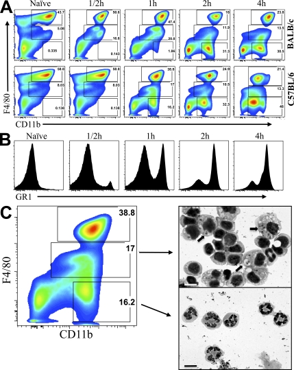
Figure 2.
Monocyte migration into the peritoneum in response to infection with L. major promastigotes. (A) Flow cytometry analysis showing the kinetics of leukocyte infiltration into the peritoneum after infection with 2 × 10⁶ L. major parasites. Three distinct populations of cells, identified by gating on CD11b and F4/80 levels, are designated by boxed areas. (B) Histograms showing GR1 expression on the F4/80IntCD11b+ monocytes between 30 min and 4 h after peritoneal infection with 2 × 10⁶ L. major. (C) Cytospin analysis of F4/80Int monocytes and F4/80Neg neutrophils after 1 h of peritoneal infection of C57BL/6 mice. Cells were gated based on FSC x SSC profile, eliminating dead cells and debris. Arrows point to L. major parasites inside cells. The numbers represent the frequency of gated cells from one experiment, and are representative of at least 4 experiments using a total of 40 mice. Bars, 10 μm.

