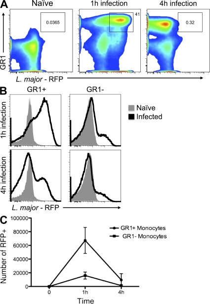
Figure 3.
The rapid killing of L. major parasites by GR1+ monocytes. (A) Flow cytometry profiles of monocytes gated on F4/80+CD11b+GR1+ monocytes at 1 and 3 h after infection with RFP-L. major. Uninfected (naive) cells show no RFP staining (left profile). (B) Histograms showing GR1+ monocytes (left) or GR1− monocytes (right) infected with RFP-L. major after 1 h (top) or 4 h (bottom) with 2 × 10⁶ RFP-L. major. (C) The number of infected (RFP-positive) F4/80+CD11b+ GR1+ monocytes (circles) and GR1− monocytes (squares) after peritoneal infection with 2 × 10⁶ L. major parasites. Cells were gated based on FSC x SSC profiles to eliminate dead cells and then separated using antibodies to F4/80 and CD11b. The number showed in dot plots represent the frequency of gated cells from one experiment and are representative of 3 independent experiments using a total of 18 mice. The numbers on the y axis represent the mean of the absolute numbers of infected monocytes, and the error bars are the SD from three independent experiments.

