Figure 4.
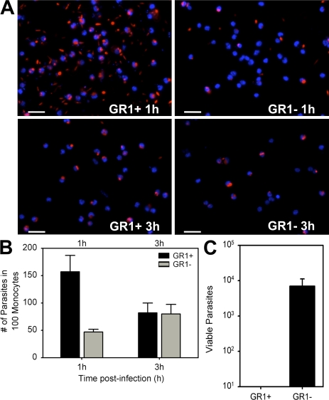
The in vitro killing of L. major by GR1+ monocytes from wild-type mice. (A) GR1+ (left) and GR1− (right) monocytes were sorted from CX3CR1GFP/+ RAG2−/− mice by GFP expression and incubated with 10:1 ratio of metacyclic RFP-L. major in vitro. Fluorescence photomicrographs of gently washed monolayers were taken after 1 h (top) or 3 h (bottom). (B) The total mean number of L. major parasites (n = 3) associated with GR1+ or GR1− monocytes after 1 or 3 h in vitro infection. (C) GR1+ and GR1− monocytes were sorted based on CX3CR1GFP expression and incubated with 5:1 ratio of L. major to monocytes in vitro. The cells were kept in culture for 3 d at 37°C without washing. After 3 d, parasite numbers were calculated by serial dilution assays. Data represent the mean ± SE of 3 independent experiments using a total of 40 mice. Bars, 50 μm.

