Figure 5.
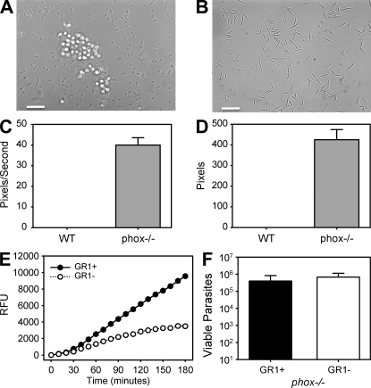
The in vitro killing of L. major parasites by GR1+ monocytes from wild-type and from phox−/− mice. (A) Phase-contrast microscopy taken on an inverted photomicroscope of sorted wild-type GR1+ monocytes showing altered morphology of unfixed metacyclic L. major promastigotes when cultivated with (A) or without (B) GR1+ monocytes for 1 h. (C and D) GR1+ monocytes were isolated from wild-type (WT) or gp91 phox−/− mice and sorted by F4/80 and GR1 expression. Monocytes were incubated with 5:1 ratio of L. major to monocytes in vitro. At 3 h the movement of extracellular promastigotes was determined using ImageJ software to calculate the mean distance (C) and velocity (D) from 20 selected parasites per culture. At least 30 mice were used in 2 independent experiments. (E) The in vitro production of ROS. The kinetics of ROS production from sorted GR1+ and GR1− infected monocytes in the presence of H2DCFDA measured by fluorometry. Data are from three independent experiments. (F) Monocytes from WT or phox−/− mice were cultivated with L. major for 3 d without washing, at which time parasite viability was determined by limiting dilution assay. Data are the mean ± SEM from three independent experiments involving at least 20 mice. Bars, 50 μm.

