Figure 7.
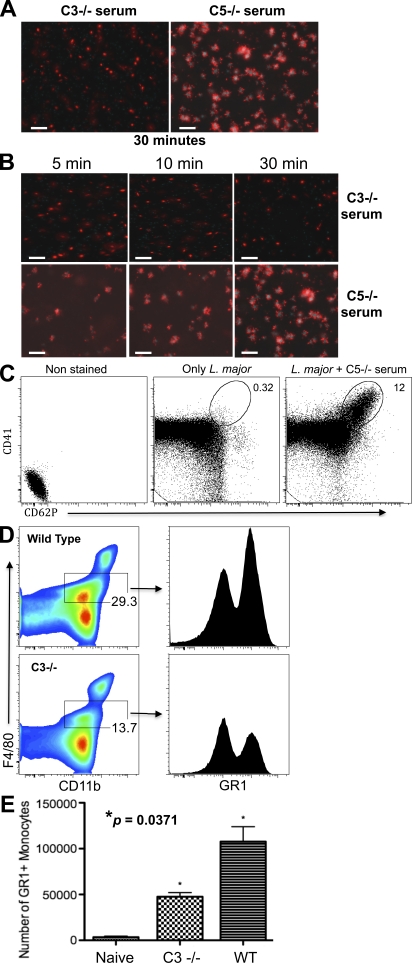
Platelet activation by L. major parasites. (A) Fluorescence photomicroscopy showing RFP-L. major parasites and CFP-labeled platelets that were incubated for 30 min in the presence of fresh nonimmune serum from C3−/− mice (left) or C5−/− mice (right). (B) 2 × 10⁷ CFP-platelets were incubated with 106 parasites for 1–30 min in the presence of serum from mice lacking C3 (C3−/−; top) or C5 (C5−/−; bottom), and then observed by fluorescence microscopy. (C) CD62P expression on platelets activated by L. major and complement. Left profile, platelets alone with no antibody; middle profile, L. major promastigotes plus platelets but no serum, stained with αCD41; right profile, L. major plus complement and platelets, stained with αCD41. (D) Monocyte recruitment into the peritoneal cavity of wild-type C57BL/6 (top) or C3−/− (bottom) mice after 4 h of infection with 2 × 10⁷ L. major parasites. The CD11b+ F4/80Int population was analyzed for GR1 expression (right profiles). (E) The total number of GR1+ monocytes migrating into the peritoneal cavity of wild-type or C3−/− mice after infection with L. major. The platelet activation experiments (A–C) were performed >5 times using at least 10 mice/experiment. The experiments using C3−/− mice (D and E) mice were performed twice using 12 mice.

