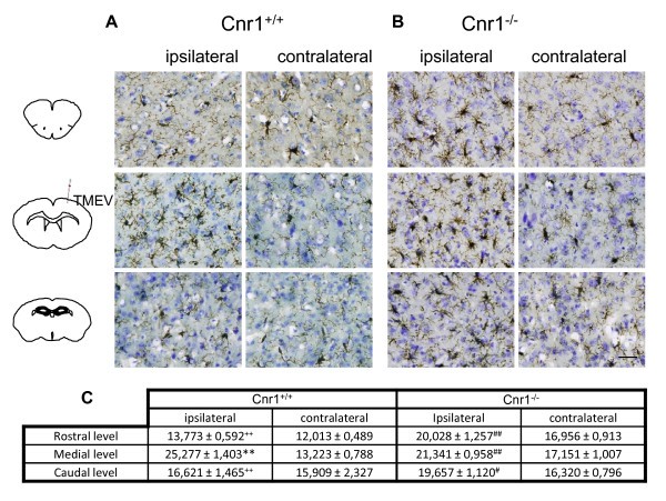
Figure 4.
CB1 deletion exacerbates microglial response against TMEV infection. Coronal brain sections (30 μm) were obtained from Cnr1+/+ TMEV-infected mice (A) or Cnr1-/- TMEV-infected mice (B), stained for CD11b with Iba-1 antibody and counterstained with toluidine blue (n = 3 for each group). To perform the analysis of microglia phenotype morphology brain tissue was studied in both hemispheres and at rostral, medial and caudal levels. (C) Quantification of percentage of area occupied by microglia per field is represented. Scale bar is 50 μm. **p < 0.01 vs. contralateral (Cnr1+/+); #p < 0.05 vs. contralateral (Cnr1-/-); ##p < 0.01 vs. contralateral (Cnr1-/-); ++p < 0.01 vs. medial level (Cnr1+/+).

