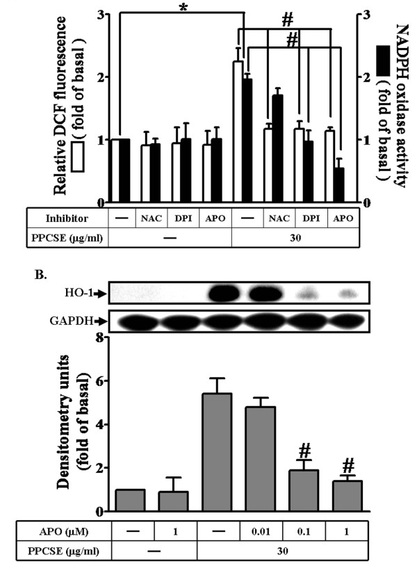
Figure 2.
PPCSE-induced HO-1 expression is mediated via NADPH oxidase-dependent ROS generation in bEnd.3 cells. A: bEnd.3 cells were labeled with DCF-DA (10 μM), and then incubated with 30 μg/ml PPCSE for 30 min with/without pretreatment of NAC (3 mM), DPI (1 μM) or APO (1 μM) for 1 h. The fluorescence intensity (relative DCF fluorescence) was measured at 495 nm excitation and 529 nm emission using a FACScan flow cytometer. The NADPH oxidase activity was measured as described in the Methods. B: Cells were pretreated with APO for 1 h, and then incubated with PPCSE for 24 h. The expression of HO-1 was determined by Western blot. Data are summarized and expressed as the mean ± SEM of three individual experiments. *P < 0.05 as compared with the cells exposed to vehicle alone. #P < 0.05 as compared with the cells exposed to PPCSE alone.

