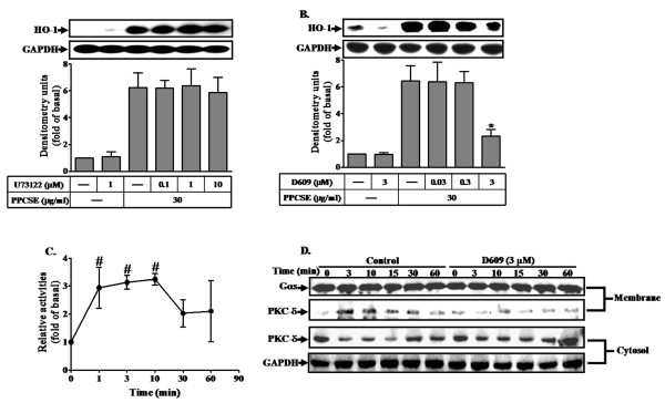
Figure 4.
PPCSE-induced HO-1 expression is mediated via a PC-PLC/PKCδ signaling in bEnd.3 cells. A, B: Cells were pretreated with the inhibitor of PI-PLC (U73122) or PC-PLC (D609) for 1 h, and then incubated with PPCSE for 24 h. The expression of HO-1 was determined by Western blot. C: Cells were treated with 30 μg/ml PPCSE for the indicated time intervals, and then the activity of Ca2+-independent PC-PLC was determined as described in the Methods. The basal level of Ca2+-independent PC-PLC was 27.4 ± 3.44 nmol/min/mg protein. D: Cells were pretreated with D609 for 1 h, and then incubated with PPCSE for the indicated time intervals. The cytosolic and membrane fractions were prepared and subjected to 12% SDS-PAGE to determine the translocation of PKCδ using an anti-PKCδ antibody. Data are summarized and expressed as the mean ± SEM of three individual experiments. *P < 0.05 as compared with the cells exposed to PPCSE alone. #P < 0.05 as compared with basal level.

