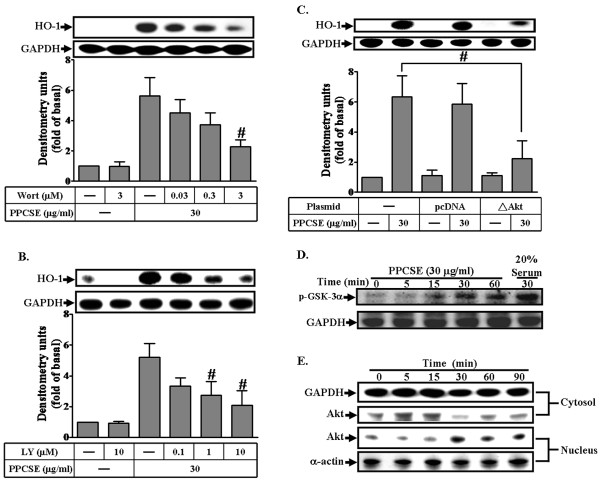
Figure 5.
Akt mediated HO-1 induction by PPCSE. A, B: Cells were pretreated with wortmannin (Wort) or LY294002 (LY) for 1 h, and then incubated with PPCSE for 24 h. The expression of HO-1 was determined by Western blot. C: Cells were transfected with a dominant negative mutant of Akt (ΔAkt), and then incubated with PPCSE for 24 h. The expression of HO-1 was determined by Western blot. D: Akt kinase activity was determined as described in the Methods. Cells were treated with 30 μg/ml PPCSE for the indicated time intervals. Phosphorylation of GSK-3α was used to determine the activity of Akt stimulated by PPCSE and analyzed by western blot. E: Time dependence of PPCSE-stimulated Akt translocation into nucleus. Cells were treated with 30 μg/ml PPCSE for the indicated time intervals. The cytosolic and nuclear fractions were analyzed by Western blot using an anti-Akt, anti-GAPDH (as a cytosol control), or anti-α-actin (as a nuclear control) antibody. A-C: Data are expressed as the mean ± SEM of three individual experiments. #P < 0.05 as compared with the cells exposed to PPCSE alone. D, E: Similar results were obtained from three independent experiments.

