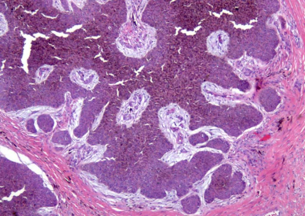
Figure 3.
High power view show invasive carcinoma cells. There is obvious cell atypia with many mitotic figures seen. The squamous cells have variable differentiation accompanied with changes in pigmentation that are aligned in a horizontal nodular pattern. Both carcinoma in situ and invasive component noted.

