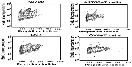
Figure 3. BrdU incorporation and propidium iodide (PI) co-staining in tumor cells.
Ovarian tumor cells, A2780 and OV4 were co-cultured for 24 h in the presence or absence of γδ T cells at the ratio of 1∶15. After co-culture, cells were pulsed with BrdU for 5 hours and PI staining was performed prior to flowcytometric analyses.

