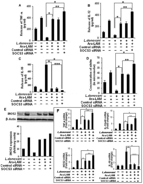
Figure 6. SOCS3 silencing significantly enhances host protective proinflammatory response generation by Ara-LAM in infected macrophages.
Peritoneal macrophages (2×10⁶ cells/mL) were transfected with control siRNA or SOCS3-specific siRNA followed by Ara-LAM treatment (for 3 hr) and Leishmania infection for 24 h and assayed for the levels of TNF- α (A), IL-12 (B), and IL-10 (C) in the culture supernatant by ELISA, as described in Methods. ELISA data are expressed as means standard deviations of values from triplicate experiments that yielded similar observations. *P<.001 compared with infected macrophages, **P<.005 compared with Ara-LAM–pretreated infected macrophages. ***<.01 compared to Ara-LAM–pretreated infected macrophages. In a separate experiment macrophages were cultured and treated as described above and assayed for the levels of extracellular NO as described in the Materials and Methods (D). *P<.001 compared with infected macrophages, **P<.005 compared with Ara-LAM–pretreated infected macrophages. In a separate set macrophages were transfected and treated with Ara-LAM as described above followed by Leishmania infection for 24 hr. Western blot analysis was performed to analyze the expression of inducible nitric oxide synthase. The blot shown is a representative of experiments performed in triplicate. Band intensities were analyzed by densitometry (E). Peritoneal macrophages were cultured and treated with Ara-LAM as described above followed by Leishmania infection for 3 hr. Changes in mRNA expression of IL-12p40, TNF- α, IL-10, NO were determined by real-time PCR analysis Results are presented as changes (n-fold) relative to uninfected control cells. The experiment was repeated 3 times, yielding similar results; data are expressed as means ± standard deviations. (F). *P<.001 compared with infected macrophages, **P<.005 compared with Ara-LAM–pretreated infected macrophages.

