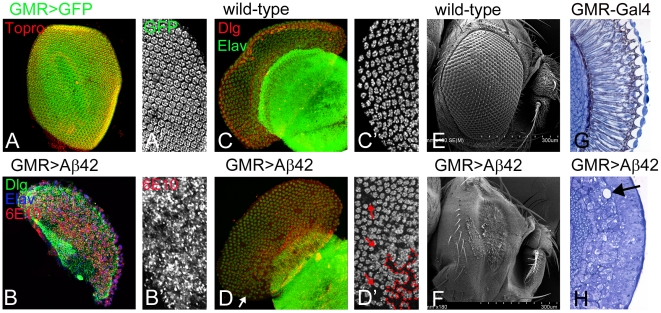
Figure 2. Aß42 accumulation in the developing pupal retina and adult eye results in neurodegeneration.
(A, C) represent normal eye development in (A) late pupal retina, and (C) the adult compound eye. Dlg (red channel) marks the membrane; 6E10 (blue channel) marks the Aß42, and Elav (green channel), a proneural marker that marks the neuronal fate. (B, D, F) Misexpression of Aß42 in the differentiating neurons of eye using a GMR-Gal4 construct (GMR>Aß42) results in onset of neurodegeneration. (C, C′) Unlike the highly organized ommatidia of wild-type pupal retina, (D, D′) the late pupal retina shows significant neuronal cell death in the eye field. By this stage, the accumulation of Aß42 plaques has resulted in distinct holes in the eye field (marked by an arrow, outline of “hole” in pupal retina marked by red dotted line). (C) Wild type adult eye with uniform arrangement of 800 unit eyes. (I) The adult eye field of GMR>Aß42 is significantly diminished and there is complete fusion of the ommatidia. (G, H) In comparison to the adult eye section of (G) wild type fly eye, (H) the GMR>Aß42 exhibits highly disorganized morphology of photoreceptors. Furthermore the retinas of GMR>Aß42 are vacuolated. This illustrates how the neurodegenerative phenotype progressively worsens as the age and dose increase over the course of fly development.

