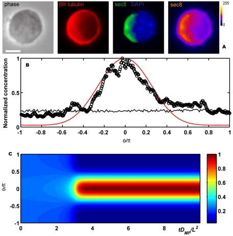
Figure 4. Symmetry breaking.
(A) Hippocampal neurons were fixed shortly after plating and immunolabeled with a neuron-specific anti- tubulin antibody (red), an anti Sec8 antibody (green) and a nuclear marker (blue). The right panel shows a pseudocolor image of the Sec8 only. Scale bar 5
  . (B) Experimental results, Sec8, (open circles) compared with numerical simulations,
  , (red line) for the round neuron shown in the inset. Initial profile (random perturbations) in black. In this case
  ,
,  ,
,  ,
,  ,
,  ,
,  and
and  . (C) Temporal evolution of the simulation shown in (B) (
 axis, membrane position;
  axis, time; color scale, normalized
  values). Results are normalized to the final maximum value of
  .
.

