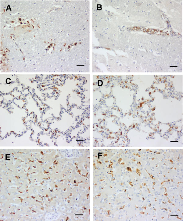
Figure 3.
CM (C). HO-1 staining of tissues from cerebral malaria cases showing physically apparent brain pathology, plus sequestered parasites present. A and B brain, C and D lung, and E and F liver. Both case 9 (A, C and E) and case 26 (B, D and F) show the brain monocyte, lung monocyte and macrophage, and Kupffer cell staining that dominate this group. Scale bar, 100 μm.

