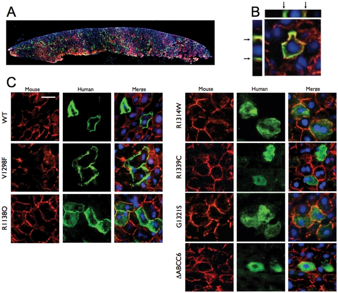
Figure 3. Intracellular localization of human ABCC6 variants expressed in vivo in mouse liver.
The human and mouse ABCC6/Abcc6 were detected on frozen sections by immunofluorescence using the M6II-31 monoclonal antibody (green) and the S-20 polyclonal antibody (red), respectively, Panel A: A low magnification image shows the overall distribution of the human WT ABCC6 in a cross-section of single liver lobe. B: The basolateral plasma membrane localization of the WT ABCC6 expressed in mouse liver hepatocytes was confirmed by immunofluorescent imaging. The Z-stack cross-section images are also shown and arrows point to the basolateral membrane. C: Localization of the human WT and mutant ABCC6. Individual channels and the merged images of the endogenous Abcc6 (red) and the human ABCC6 variants (green) are shown. The scale bar represents 50 µm.

