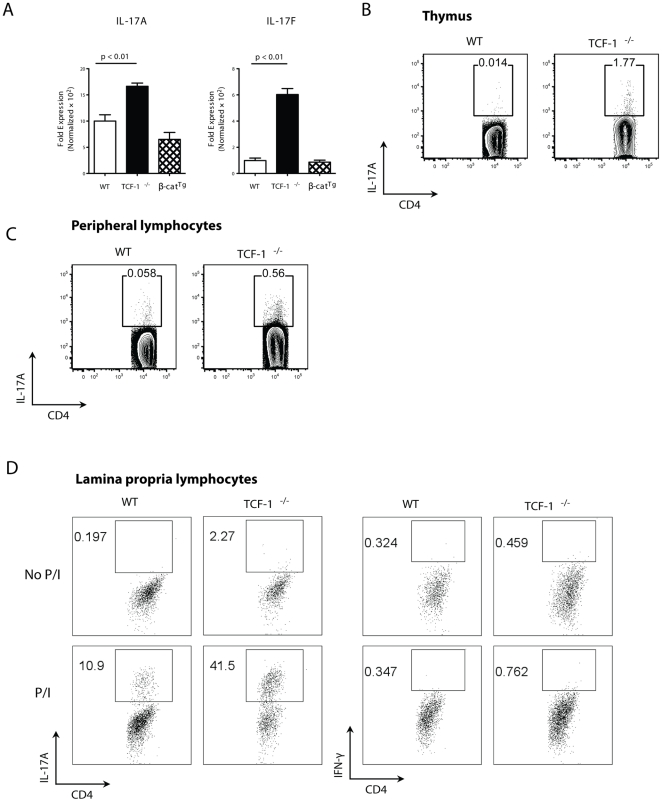
Figure 1. TCF-1 is required to repress the IL-17 gene during T cell development.
(A) Increased expression of IL-17 mRNA in TCF-1-/- but not β-catTg thymocytes. Levels of IL-17A and IL-17F mRNAs from WT, TCF-1-/- or β-catTg thymocytes, as determined by qRT-PCR; n = 4; error bars indicate ±SEM. (B-C) Increase in natural Th17 cells in TCF-1-/- mice. T cells from thymus (B) or peripheral lymphocytes (C) of WT or TCF-1-/- mice were stimulated with PMA and ionomycin for 4 h immediately after isolation, and the percentage of natural Th17 gated on CD4+TCR-β+ cells was detected by intracellular staining for IL-17. Shown contour plots are representative of three independent experiments. (D) Increased IL-17 but not IFNγ producing T cells in lamina propria lymphocytes. Lamina propria lymphocytes from WT and TCF-1-/- mice were either not treated (top panels) or stimulated with PMA/Ionomycin (bottom panels). IL-17 and IFNγ producing cells were detected by flow cytometry.

