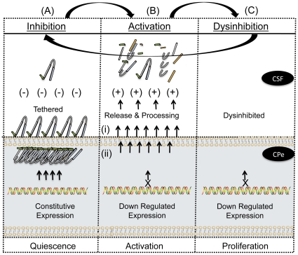
Figure 5. A biphasic model for augurin activity and CNS dysinhibition.
Normal choroid plexus epithelia express Ecrg4 and contain significant 14 kDa augurin protein that is in an inhibitory conformation. The unprocessed peptide is contained in 3 compartments (1) intracellular vesicles (i.e. punctate intracellular staining in Figure 1) (2) near the ventricular cell surface (i.e. polarized apical staining of cells in Figure 1 and (3) tethered at the cell surface (i.e. Figure 1B). Upon injury, the sudden release and processing at the cell surface is followed by release from intracellular stores as a “panic signal” to indicate systemic injury. The initial pro-inflammatory response then shuts down Ecrg4 gene expression for at least 1 to 3 days post-injury. Therefore, during the initial phase of injury, its normal, constitutively inhibitory functions are absent and so repair cells proliferate. As gene expression returns, quiescence is restored and homeostasis re-established.

