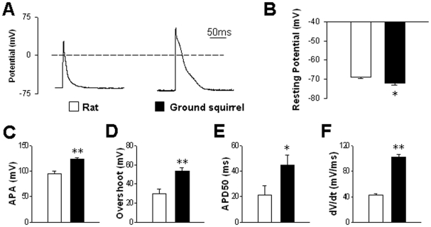
Figure 3. Action potentials evoked by current pulses.
(A) Typical examples of action potentials in ventricular myocytes from rats (left) and ground squirrels (right). (B) Resting membrane potentials were compared between rats (n = 18 cells from 7 animals) and ground squirrels (n = 21 cells from 7 animals). (C) Action potential amplitude (APA), (D) overshoot, (E) half-repolarization duration (APD50) and (F) maximum dV/dt of depolarization were compared between rats (n = 7 cells from 5 animals) and ground squirrels (n = 6 cells from 5 animals). *P<0.05 and **P<0.01.

