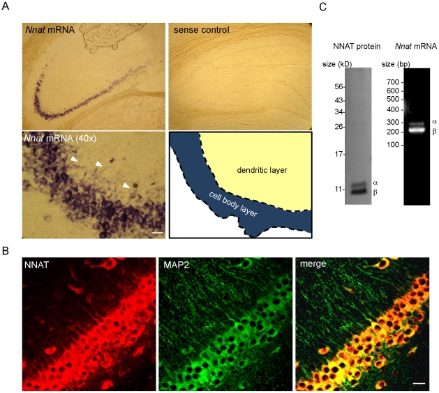
Figure 1. Neuronatin is expressed in the P21 rat hippocampus.
(A) Nnat in situ hybridization (ISH) in hippocampus visualized using alkaline phosphatase. Nnat mRNA is present in hippocampal regions CA2 and 3. Arrowheads denote dendritic localization. Scale bar: 20 µm. Schematic of cell body and dendritic layer boundary in 40× image is shown (bottom right panel). (B) NNAT immunofluorescent staining in the CA2 region of hippocampus showing dendritic localization, NNAT (red), MAP2 (green). Scale bar: 20 µm. (C) left, Western blot for NNAT and right, RT-PCR for Nnat mRNA using rat hippocampal tissue showing both α and β isoforms.

