
Figure 6.
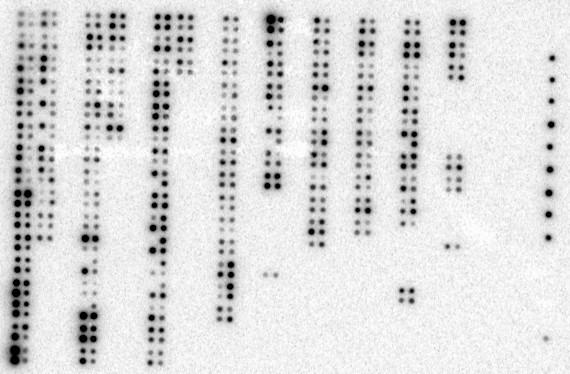
Expression of CUL-5 in normal tissues versus matched tumor tissues. The cancer profiling array was hybribized with the 32P-labeled (2 × 10⁷ cpm) 674-bp CUL-5 cDNA for 20 hrs. The results shown represent the 2-day exposure to the phosphor screen. The location of individual samples on the array is shown in Figure 5.