
Figure 7.
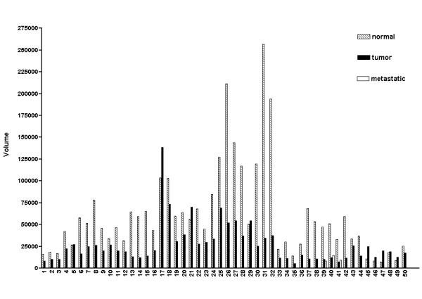
Expression of CUL-5 in the 50 cases of matched normal breast tissue and breast tumor tissue. The data in this figure was derived from quantification of the data in Figure 6 using a PhosphorImager and ImageQuant software, and the background adjusted volumes are shown for each sample. 41 of the 50 cases of breast tissue (82%) exhibited a decrease in CUL-5 expression in the tumor tissue versus the matched normal tissue. Three of the 50 cases had a matched metastatic sample (cases 39, 40, and 41), and the level of CUL-5 expression in the metastatic samples was similar to that seen in the associated tumor tissues.