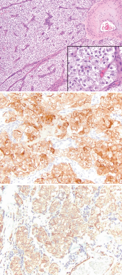
Fig. 1.
Nasal PEComa (Case 1) comprised of sheets and nests of polygonal cells associated with a prominent vascular stroma. The cells are cytologically bland with abundant clear to eosinophilic granular cytoplasm (top; H&E 100×; inset 400×). The tumor was strongly positive for Melan-A (middle; 200×) and calponin (bottom; 200×)

