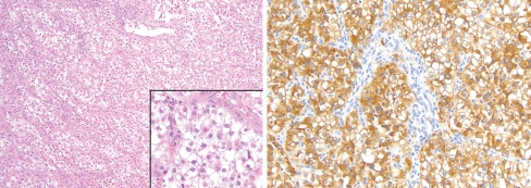
Fig. 3.
The laryngeal PEComa (Case 3) was histologically identical to Case 1, with characteristic polygonal cells and a delicate vascular stroma (inset), but showed foci with the characteristic perivascular distribution of tumor cells as seen in the upper field (left; H&E 100×; inset 400×). The tumor was strongly positive for HMB45 (right; 200×) and patchy SMA staining

