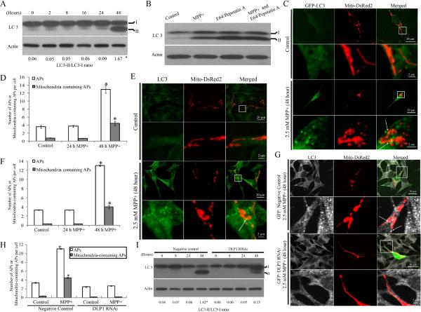
Figure 7. MPP+-induced mitochondrial fragmentation was prerequisite for MPP+-induced Autophagy/mitophagy.
(A) Representative immunoblot of LC3 and quantification of LC3-II/LC3-I ratio in SH-SY5Y cells treated with 2.5 mM MPP+ for various periods of time. (B) SH-SY5Y cells were treated with 2.5mM MPP+ in the absence or presence of E64 (10μg/ml) and pepstatin A (10μg/ml) to inhibit lysosomal degradation. After 48 h, cell lysates were analyzed for LC3 by immunoblot. (C,D) To visualize autophagosome formation, SH-SY5Y cells were transiently co-transfected with GFP-LC3 and mito-DsRed2. Two days after transfection, cells were treated with 2.5 mM MPP+ for 24 or 48 h. (C) and (D) are representative immunofluorescence pictures and quantification of autophagosome (AP) in cells transfected with GFP-LC3 and treated with 2.5 mM MPP+ for 24 or 48 h. At least 50 cells were analyzed in each experiment. (E,F) Autophagosome formation was also assayed by direct immunostaining of LC3 in SH-SY5Y cells fixed after treatment of 2.5 mM MPP+ for 24 or 48 h (E) and number of APs was quantified (F). At least 50 cells were analyzed in each experiment. (G,H) To study the effect of DLP1 reduction on MPP+ induced autophagy/mitophagy, SH-SY5Y cells were co-transfected with empty/negative control vectors or DLP1 RNAi constructs and mito-DsRed2. Two days after transfection, cells were treated with MPP+ for 48 h, fixed and stained by LC3 (G) and the number of APs was quantified (H). At least 50 cells were analyzed in each experiment. (I) Representative immunoblot and quantification analysis of LC3 in DLP1 RNAi SH-SY5Y cells after treatment of 2.5 mM MPP+ for various periods of time. Equal protein amounts (10 μg) were loaded and actin was used as an internal loading control. All experiments were repeated three times. (*p<0.05, when compared with non-treated cells; student-t-test).

