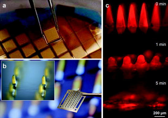
Fig. 3.

Solid microneedle patches. a Arrays of solid silicon microneedles coated with gold. (Courtesy of University of Queensland). b Array of solid stainless steel microneedles coated with yellow dye. Each 12 mm by 12 mm device contains 50 microneedles measuring 700 μm tall. Inset shows magnified view of two coated microneedles (Courtesy of Georgia Institute of Technology). c Dissolving microneedles shown intact before insertion into skin, partially dissolved 1 min after insertion into skin and fully dissolved 5 min after insertion into skin (Reproduced from (Sullivan et al. 2010); Courtesy of Georgia Institute of Technology)
