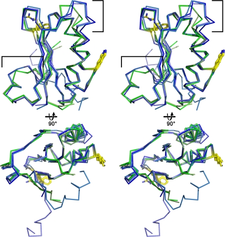
Fig. 7.
Stereo images of superpositioned single-domain BMC monomers from the β- (blue shades) and α- (green shades) carboxysomes. The upper pair is viewed from the convex side of the protein, whereas the bottom view is rotated clockwise 90° about the x-axis from the upper view. One pore residue (Arg from CcmK4, Lys from CcmK1 and CcmK2, Phe from CsoS1A and CsoS1C) and the conserved Lys found at the edge of the hexamer are shown in yellow sticks. The regions flanked by brackets are those that display the largest structural differences between the Cso and CcmK type shell proteins

