
Figure 6.
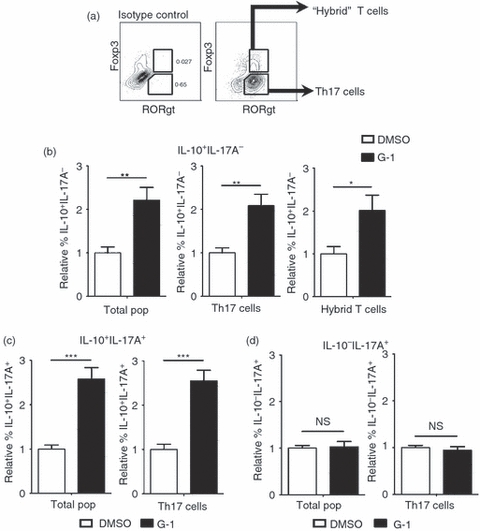
G-1 induces interleukin-10 (IL-10) production within the hybrid T-cell population. CD4+ CD62Lhi naive CD4+ T cells were collected by FACS from Foxp3egfp mice and cultured for 4 days ex vivo with anti-CD3/28 + IL-6 + transforming growth factor-β (TGF-β) in the presence of 100 nm G-1 (black bars) or DMSO (white bars). Cells were collected and stained for intracelluar IL-10, IL-17A, and RORγt, and analysed by flow cytometry. Cells that were Foxp3+ RORγt+ were designated as hybrid T cells, whereas those that were Foxp3− RORγt+ were designated as T helper type 17 (Th17) cells. (a) Gating logic to determine hybrid T-cell and Th17 populations. (b–d) Graphs represent summary of data from three independent experiments showing the relative percentage of (b) IL-10+ IL-17A−, (c) IL-10+ IL-17A+, and (d) IL-10− IL-17A+ populations. P-values determined by Student's t-test; *P < 0·05; **P < 0·005; ***P < 0·0005. Error bars = SEM; NS, not significant.